Medical demonstration model of lumbar intervertebral disc herniation in white PVC material measuring 11x10x7.6 cm showing spinal disc anatomy and pathology for medical education and training purposes.


Medical demonstration model of lumbar intervertebral disc herniation in white PVC material measuring 11x10x7.6 cm showing spinal disc anatomy and pathology for medical education and training purposes.
White PVC lumbar intervertebral disc herniation model measuring 11x10x7.6 cm for medical education

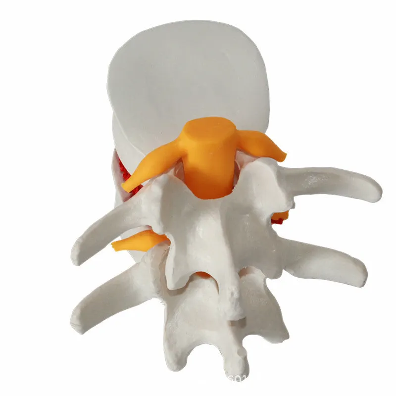
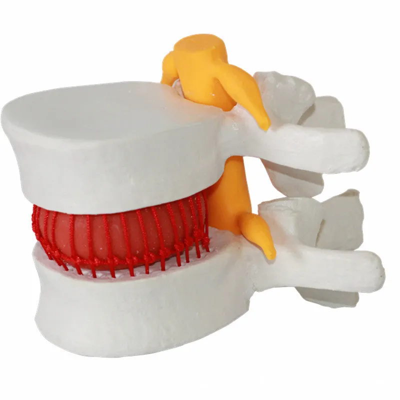
This anatomical demonstration model provides healthcare educators and students with a tangible representation of lumbar intervertebral disc pathology. Measuring 11 cm in length, 10 cm in width, and 7.6 cm in height, the three-dimensional format allows for detailed examination of disc herniation characteristics that two-dimensional illustrations cannot convey. The white PVC construction offers clear contrast for identifying anatomical structures, while the 0.22 kg weight makes it portable between classroom settings, clinical environments, and training sessions. This model serves as a practical tool for demonstrating spinal anatomy to both medical professionals and patients during educational discussions.
Features and Construction
The lumbar disc herniation model is designed specifically for educational visualisation of spinal pathology. Its construction focuses on anatomical accuracy within practical handling parameters, balancing detail with durability for classroom use.
Material and Build
Constructed from PVC (polyvinyl chloride), this model provides a stable base material that maintains its shape during repeated handling. The white colouration offers optimal contrast for visualising anatomical details, particularly useful when demonstrating disc herniation to groups. The material surface allows clear observation of pathology without excessive reflection that might obscure details under classroom lighting conditions.
Size and Practical Fit
With dimensions of 11 cm in length, 10 cm in width, and 7.6 cm in height, this model provides sufficient detail for close examination while remaining manageable for single-handed demonstration. The 0.22 kg weight ensures easy portability between teaching locations, and the compact size allows storage in standard educational supply cabinets. The packaging measures 11.5x11x8 cm, providing secure transit protection while minimising storage space requirements.
Uses and Placement

This demonstration model supports various educational scenarios where three-dimensional representation enhances understanding of spinal anatomy and pathology.
Event or Professional Use
In medical education settings, the model serves as a visual aid during lectures on spinal disorders, orthopaedic assessments, or neurology modules. Healthcare professionals may use it during patient consultations to explain disc herniation mechanics, surgical procedures, or conservative treatment approaches. The clear white presentation makes it suitable for photography or video recording for digital educational materials.
Everyday Home Use
Medical students can utilise this model for self-study and revision of spinal anatomy, particularly when preparing for practical examinations. The durable PVC construction withstands regular handling during study sessions, and the compact size allows placement on desks or bookshelves alongside other educational materials.
Benefits and Buying Value

This anatomical model provides practical advantages for medical education through its specific focus and construction approach.
Reuse and Low Maintenance
The PVC material requires minimal maintenance beyond occasional cleaning with a damp cloth, making it suitable for repeated use across multiple academic terms or training sessions. The durable construction maintains anatomical accuracy despite regular handling, ensuring consistent educational value over time without degradation of key features.
Why Choose This Product
This model offers specific focus on lumbar intervertebral disc herniation rather than general spinal anatomy, providing targeted educational value for this common pathology. The combination of practical dimensions (11x10x7.6 cm) and lightweight construction (0.22 kg) balances detail with portability, while the white PVC presentation ensures clear visibility of anatomical features in various lighting conditions common to educational environments.